
¿QUE ES?
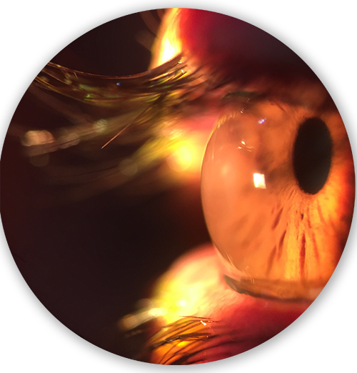
El queratocono es una ectasia corneal progresiva, bilateral, crónica y asimétrica(1)(2)(3). Se caracteriza por un adelgazamiento y protrusión corneal localizado, que cursa con miopía y astigmatismo irregular afectando significativamente la calidad visual(4)(5)(6)(7).
Aparece típicamente durante la pubertad (1)(2)(4)(8), y progresa hacia la tercera(1) y cuarta década de la vida (4)(8)(9).
El queratocono es la ectasia primaria más común, su incidencia se estimaba en 2 casos por 100.00 por año(10), sin embargo, en un estudio reciente se ha evidenciado que la incidencia anual es de 13.3 casos por 100.000 y la prevalencia de 265 casos por 100.000, lo que representa un aumento en los casos de queratocono(1)(8), debido probablemente a un mayor conocimiento sobre la enfermedad y a una mejoría en las técnicas de diagnóstico como la topografía y tomografía corneal, que han permitido detectar la ectasia incluso en estadios más tempranos (1)(11).
ETIOLOGIA
La etiología del queratocono aún no es comprendida del todo(1)(5)(12), sin embargo, se ha establecido que es multifactorial(1)(2)(3): factores tales como alergias, frotamiento ocular (13)(14), mecanismos genéticos, inflamación y estrés oxidativo juegan un papel en la patogénesis(15). Su aparición también se ha asociado a trauma repetitivo con lentes de contacto(1)(16)(17)(18).
TRATAMIENTO
El manejo clínico del queratocono depende de su severidad(19), y se puede dividir en tratamiento quirúrgico y no quirúrgico(11). En su etapa leve, el manejo se inicia con gafas o lentes de contacto blandos(2)(4), mientras que en los casos más avanzados la mejoría visual se puede lograr con varios tipos de lentes de contacto (LC) como rígidos gas permeables (RGP), blandos especiales para queratocono, híbridos, Piggy Back, o esclerales(20). Los RGP son la opción de tratamiento más común y exitosa, especialmente en queratoconos leves a moderados(21)(22).
El objetivo clínico del uso de lentes de contacto gas permeables es proporcionar mejor visión, comodidad y máximo tiempo de uso posible, minimizando cualquier efecto adverso sobre la fisiología corneal(23).

BIBLIOGRAFIAS
Mas-Tur V, MacGregor C, Jayaswal R, O’Brart DPS, Maycock NJR. A review of keratoconus: diagnosis, pathophysiology and genetics. Surv Ophthalmol. 2017.
Naderan M, Shoar S, Rezagholizadeh F, Zolfaghari M, Naderan M. Characteristics and associations of keratoconus patients. Contact Lens Anterior Eye. 2015;38(3):199–205.
Muftuoglu O, Ayar O, Ozulken K, Ozyol E, Akinci A. Posterior corneal elevation and back difference corneal elevation in diagnosing forme fruste keratoconus in the fellow eyes of unilateral keratoconus patients. J Cataract Refract Surg. 2013;39(9):1348–57.
Romero-Jimenez M, Santodomingo-Rubido J, Wolffsohn JS. Keratoconus: A review. Contact Lens Anterior Eye. 2010;33(4):157–66.
Galvis V, Sherwin T, Tello A, Merayo J, Barrera R, Acera A. Keratoconus: an inflammatory disorder? Eye. 2015;29(7):843–59.
Lema I, Durán JA. Inflammatory molecules in the tears of patients with keratoconus. Ophthalmology. 2005;112(4):654–9.
Mandathara PS, Fatima M, Taureen S, Dumpati S, Ali MH, Rathi V. RGP contact lens fitting in keratoconus using FITSCAN technology. Cont Lens Anterior Eye. 2013 Jun;36(3):126–9.
Godefrooij DA, de Wit GA, Uiterwaal CS, Imhof SM, Wisse RPL. Age-specific Incidence and Prevalence of Keratoconus: A Nationwide Registration Study. Am J Ophthalmol. 2017;175:169–72.
Ortiz-toquero S, Rodriguez G, Juan V De, Martin R. Contact Lens and Anterior Eye New web-based algorithm to improve rigid gas permeable contact lens fi tting in keratoconus. 2017;40:143–50.
Rabinowitz YS. Keratoconus. Surv Ophthalmol. 1998;42(4):297–319. Available from:
Gomes JAP, Tan D, Rapuano CJ, Belin MW, Ambrósio R, Guell JL, et al. Global Consensus on Keratoconus and Ectatic Diseases. Cornea. 2015 Apr [cited 2017 Jan 10];34(4):359–69.
Bilgin LK, Yılmaz Ş, Araz B, Yüksel SB, Sezen T. 30 years of contact lens prescribing for keratoconic patients in Turkey. Contact Lens Anterior Eye. 2009;32(1):16–21.
Carracedo G, González-Méijome JM, Martín-Gil A, Carballo J, Pintor J. The influence of rigid gas permeable lens wear on the concentrations of dinucleotides in tears and the effect on dry eye signs and symptoms in keratoconus. Contact Lens Anterior Eye. 2016;39(5):375–9.
Nejabat M, Khalili MR, Dehghani C. Cone location and correction of keratoconus with rigid gas-permeable contact lenses. Contact Lens Anterior Eye. 2012;35(1):17–21.
Toprak I, Yaylalı V, Yildirim C. A combination of topographic and pachymetric parameters in keratoconus diagnosis. Contact Lens Anterior Eye. 2015;38(5):357–62.
Balasubramanian SA, Pye DC, Willcox MDP. Levels of lactoferrin, secretory IgA and serum albumin in the tear film of people with keratoconus. Exp Eye Res. 2012;96(1):132–7.
Davidson AE, Hayes S, Hardcastle AJ, Tuft SJ. The pathogenesis of keratoconus. Eye. 2014;28(2):189–95.
Dogru M, Karakaya H, Özçetin H, Ertürk H, Yücel A, Özmen A, et al. Tear function and ocular surface changes in keratoconus. Ophthalmology. 2003;110(6):1110–8.
Bhatoa NS, Hau S, Ehrlich DP. A comparison of a topography-based rigid gas permeable contact lens design with a conventionally fitted lens in patients with keratoconus. Contact Lens Anterior Eye. 2010;33(3):128–35.
Moschos MM, Nitoda E, Georgoudis P, Balidis M, Karageorgiadis E, Kozeis N. Contact Lenses for Keratoconus- Current Practice. Open Ophthalmol J. 2017;11:241–51.
Ghosh S, Mutalib HA, Sharanjeet-Kaur, Ghoshal R, Retnasabapathy S. Effects of contact lens wearing on keratoconus: a confocal microscopy observation. Int J Ophthalmol. 2017;10(2):228–34.
Romero-Jiménez M, Santodomingo-Rubido J, Flores-Rodríguez P, González-Méijome JM. Short-term corneal changes with gas-permeable contact lens wear in keratoconus subjects: A comparison of two fitting approaches. J Optom. 2015;8(1):48–55.
Zadnik K, T.. BJ, Steger-May K, Edrington T, McMahon TT, Gordon MO. Comparison of Flat and Steep Rigid Contact Lens Fitting Methods in Keratoconus. Optom Vis Sci. 2005;82(12):1014–21.